
Mechanisms Behind Bronchogen’s Anti-Inflammatory Effects Researchers study Bronchogen as a peptide complex that influences inflammation at the cellular level in
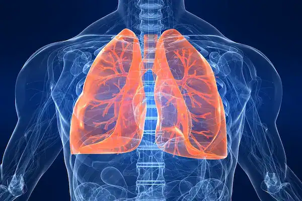
Researchers study the bronchogen peptide in laboratory models to examine its role in lung and bronchial tissue maintenance. The peptide interacts with bronchial epithelial cells and influences cellular organization during tissue stress. Research shows that bronchogen supports signaling pathways linked to airway structure and gene activity involved in tissue stability. These effects make the peptide relevant to experimental studies of cellular regulation in lung tissue.
Research also shows that the bronchogen peptide influences inflammatory activity in lung tissue models. Scientists observe reduced expression of pro-inflammatory markers, including TNF-α, alongside improved tissue structure in damaged bronchial samples. These findings position the peptide as a key focus in lung tissue research centered on cellular responses and airway integrity.
Understanding this broad tissue-level role creates a foundation for examining how bronchogen affects more specific lung components, starting with airway cells that form the first structural and functional barrier.
Explore Bronchogen Peptide from Direct Peptides United Kingdom, a lung-targeting regulatory peptide that supports epithelial stability, airway integrity, and balanced cellular activity.
The bronchogen peptide changes airway cell behavior by preserving normal epithelial layer composition during experimental stress. Airway cells exposed to the peptide maintain balanced proportions of functional cell types, including ciliated cells that support mucociliary clearance. This effect limits abnormal epithelial remodeling and helps airway cell layers retain organized structure in research models.
The peptide also alters airway cell responses to local stress signals. Treated airway cells show reduced inflammatory output and stronger support for mucosal barrier activity. These changes improve airway cell stability and local defense responses without altering overall tissue architecture, making the peptide a focus in airway cell outcome studies.
Because airway cell behavior directly influences inflammatory responses, examining how bronchogen affects lung inflammation provides further insight into its regulatory scope.
Explore Peptide Supplies at Direct Peptides United Kingdom for all your reconstitution requirements.
Research also shows that bronchogen normalizes pro-inflammatory cytokine profiles and increases secretory immunoglobulin A levels in bronchoalveolar lavage fluid (BALF). Secretory immunoglobulin A supports control of inflammatory responses at the airway surface through mucosal immune regulation. Together, these findings show that bronchogen attenuates excessive inflammatory activity in experimental lung inflammation models while supporting regulated local immune responses.
Beyond inflammation, deeper insight emerges by examining molecular-level changes that guide long-term lung tissue behavior.
Research models show that bronchogen alters the expression of genes involved in airway cell differentiation and epithelial identity. Lung tissue exposed to bronchogen demonstrates changes in transcription factors such as Nkx2.1 and SCGB1A1, which regulate epithelial specialization and secretory cell behavior. Additional shifts occur in regulatory genes like FoxA1 and FoxA2, both of which influence epithelial maintenance and lung tissue organization.
Experimental models also show bronchogen-associated changes in genes related to mucus regulation (MUC4, MUC5AC) and cellular survival markers, including Ki67 and Mcl-1. These gene expression patterns align with controlled epithelial turnover and reduced abnormal cellular activity, indicating that bronchogen directs gene regulation toward stable lung tissue behavior rather than uncontrolled remodeling.
While bronchogen remains central, lung tissue research often benefits from studying additional peptides that address complementary regulatory pathways.
Alongside the bronchogen peptide, researchers examine other regulatory peptides to broaden understanding of lung tissue behavior in experimental models. VIP and B7-33 receive attention in lung research because they influence airway signaling and tissue regulation through pathways that differ from those associated with bronchogen.
These peptides complement bronchogen peptide research by addressing separate aspects of lung tissue regulation. VIP focuses on airway responsiveness and inflammatory signaling, while B7-33 targets structural remodeling processes linked to tissue integrity. Together, they help expand lung tissue research by covering functional and structural regulation within controlled research.
Lung Tissue Research Peptides Comparison
| Research Peptide | Target Receptor / Pathway | Primary Biological Effect |
|---|---|---|
| Bronchogen | NKX2.1 / SCGB1A1 gene pathways | Modulates epithelial differentiation and regulated airway cellular behavior |
| VIP | VPAC1 / VPAC2 receptors | Promotes airway smooth muscle relaxation and immune modulation |
| B7-33 | RXFP1 receptor | Attenuates fibrotic tissue formation and supports collagen remodeling |
Check out VIP Peptide from Direct Peptides United Kingdom, a neuropeptide that helps reduce airway constriction and inflammation for improved lung function and breathing.
Vasoactive intestinal peptide (VIP) acts directly on the lung’s airway and vascular systems by binding to specific receptors expressed on airway smooth muscle and immune cells in lung tissue. VIP triggers bronchodilation and vasodilation, helping airway and pulmonary blood vessel smooth muscles relax in experimental lung models. This effect supports airway patency and normal airflow in controlled settings.
Studies also show that VIP reduces inflammatory signals in lung tissue and limits inflammatory cell infiltration in experimental lung injury and chronic airway conditions. VIP’s regulatory actions include inhibition of pro-inflammatory activity and modulation of local immune responses, contributing to more balanced lung tissue signaling in research.
Structural remodeling represents another important dimension of lung tissue research, particularly in conditions associated with fibrosis.

B7-33 is a single-chain derivative of the relaxin-2 peptide that engages the relaxin receptor RXFP1 and shows anti-fibrotic effects in experimental models of organ fibrosis. In preclinical rodent models of chronic airway injury and allergic airway disease, B7-33 significantly reduced collagen deposition and thickening of airway walls, both key markers of fibrosis, and partially improved airway responsiveness.
Research shows B7-33 promotes activity of enzymes like matrix metalloproteinase-2, which facilitate collagen breakdown and help remodel fibrotic tissue, and activates signaling pathways such as ERK1/2 associated with tissue repair in fibroblast-rich environments. These outcomes indicate that B7-33 influences lung tissue structure by reducing fibrotic buildup and supporting tissue remodeling in controlled research.
Taken together, these research findings point toward broader possibilities for lung-focused peptide studies.
Shop B7-33 Peptide from Direct Peptides United Kingdom, a relaxin-derived peptide that promotes anti-fibrotic activity and supports lung tissue remodeling in experimental models.
Bronchogen peptide research continues to guide lung tissue studies by helping researchers examine regulatory signaling, tissue stability, and adaptive responses in controlled models. Its focused activity makes it a strong reference point for understanding how lung tissue responds to sustained stress and repair cues without expanding beyond experimental boundaries.
Looking ahead, researchers are likely to study bronchogen alongside peptides such as VIP and B7-33 to better understand coordinated lung tissue regulation. This combined research approach may clarify how airway function, immune balance, and structural remodeling interact within lung tissue research frameworks as experimental methods continue to advance.
[1] Raby KL, Michaeloudes C, Tonkin J, Chung KF, et al. Mechanisms of airway epithelial injury and abnormal repair in asthma and COPD. Front Immunol. 2023 Jul 13;14:1201658.
[2] Menk M, Graw JA, Steinkraus H, Haefen Cv, et al. Characterization of inflammation in a rat model of acute lung injury after repeated pulmonary lavage. Exp Lung Res. 2015;41(8):466-76.
[3] Kuzubova NA, Lebedeva ES, Dvorakovskaya IV, Surkova EA, et al. Modulating Effect of Peptide Therapy on the Morphofunctional State of Bronchial Epithelium in Rats with Obstructive Lung Pathology. Bull Exp Biol Med. 2015 Sep;159(5):685-8.
[4] Wu D, Lee D, Sung YK. Prospect of vasoactive intestinal peptide therapy for COPD/PAH and asthma: a review. Respir Res. 2011 Apr 11;12(1):45.
[5] Hossain MA, Kocan M, Yao ST, Royce SG, et al. A single-chain derivative of the relaxin hormone is a functionally selective agonist of the G protein-coupled receptor, RXFP1. Chem Sci. 2016 Jun 1;7(6):3805-3819.
Shop ALL Peptide Vials from Direct Peptides United Kingdom today, your trusted supplier of premium clinical grade peptides online.
Bronchogen Peptide Vial
Price range: £41.50 through £50.48 Select options This product has multiple variants. The options may be chosen on the product page
B7-33 2mg Pre-Mixed Peptide
Price range: £22.55 through £60.88 Select options This product has multiple variants. The options may be chosen on the product page
Thymosin Alpha-1 LL-37 VIP Peptide Stack
Original price was: £111.88.£100.69Current price is: £100.69. Add to cart
VIP Nasal Spray
Price range: £26.24 through £47.48 Select options This product has multiple variants. The options may be chosen on the product pageMechanisms Behind Bronchogen’s Anti-Inflammatory Effects Researchers study Bronchogen as a peptide complex that influences inflammation at the cellular level in

The Role of AMPK Activators United Kingdom in Boosting Cellular Energy AMPK, or AMP activated protein kinase, works as a

Why Does PEG MGF Peptide United Kingdom Last Longer Than Regular MGF? The PEG-MGF peptide lasts longer in research environments

How Can Pegylated MGF United Kingdom Reduce Cortisol? Cortisol, often called the “stress hormone,” plays a crucial role in metabolism,